

Alzheimer’s disease, Parkinson’s disease, type 2 diabetes, and so many more. These days more and more discussions around diseases mention these sticky proteins called “amyloids,” but what really are amyloids, and why do we call them that? Even many of the scientists who study amyloids may not have an answer, but to really answer the question, we must first go back almost 400 years.
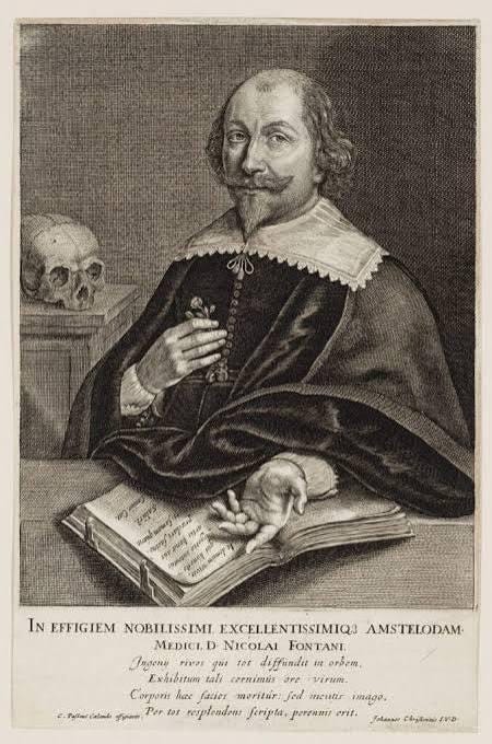
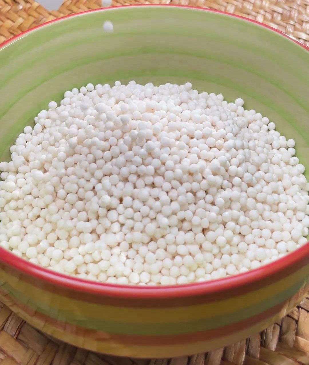
The University of Leyden was founded in 1575 as an institute dedicated to science and medicine, but also to arts like poetry and theater. It was here a physician named Nicholas Fontanus first discovered amyloids in 1639. Fontanus wasn’t just a physician; he was a poet, a playwright, and a visionary. When he saw a young man die of jaundice, he knew there had to be an answer for what happened. He conducted an autopsy and reported “large white stones” in the spleen of the patient, comparing them to the grains of a sago palm1,2. However, soon after, his discovery was corroborated by Thomas Bartholin, a professor at the University of Copenhagen, Denmark, in 1654 when he published his Historiarum Anatomicarum Rariorum. Bartholin had discovered a similar phenomenon while performing an autopsy of a woman whose spleen was so hard it could barely be cut, describing the process as cutting through “spongy timbers”3. In 1722 Jeremiah Wainewright reported a similar phenomenon in the liver of an autopsied patient with a swollen neck. He had expected the patient to have succumbed to thyroid cancer, only to discover their liver had been swollen nearly 2-3 times the size of a normal liver. He reported the liver was full of a “pale gelatinous material,” an observation further confirmed in 1789 by Antoine Portal, a French physician and medical historian who noticed a similar liver in an elderly woman. Although, when Portal observed another duplicate case in a young boy, he decided to heat up the “lard-like” substance from the boy’s liver and noticed rather than deteriorate, it actually got harder, like boiling an egg 2,4.
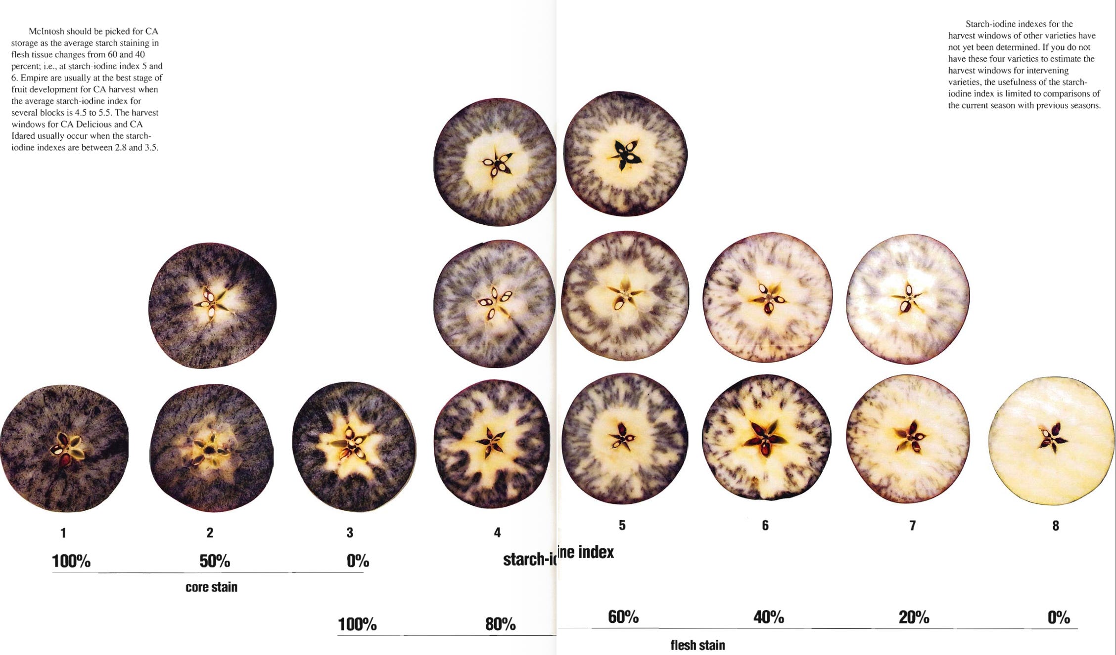
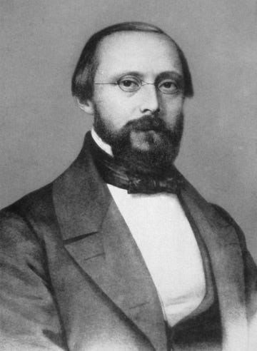
It wouldn’t be until the mid-1800s, after years of this mysterious diagnosis popping up all over the globe, that Rudolf Virchow would enter the arena. Virchow, who is often considered the “father of modern pathology,” was the first to claim “people don’t get sick, collections of cells do”5. During his time as chair of pathologic anatomy at the University of Bavaria in Würzburg, he claimed these “stones” or “plaques” were actually starch build-ups, with some peers suggesting cellulose, the indigestible plant product, as a culprit. This hypothesis was supported by using iodine as a stain (often with sulfuric acid), which showed a distinct color change when added to the mysterious plaques, much like the reaction of iodine when added to starches, a common experiment for kids in biology class. What Virchow and his peers didn’t know at that time was that complex protein structures and aggregates can interact with iodine in a similar way that starches do, causing the same color change6–8. This discovery of starch build-ups in his patients’ organs is what ultimately led Virchow to begin using the term “amyloid” in reference to the deposits he witnessed while studying the “corpora amylacea,” amyloid deposits often associated with tumors and tissue damage.
Virchow would continue to use the term to explain the spleen and liver conditions previously mentioned in medical history as examples of amyloidosis 2,3,9. The term “amyloid” itself wasn’t coined by a physician or a biochemist, but instead by a botanist, Matthias Schleiden. In 1838, Schleiden coined the term “amyloid” to describe typical starch-filled areas of plants and was only used by Virchow to describe the protein aggregates in the body because of his mistaken belief they were composed of starch 2,3,6,8,10. Therefore, the term “amyloid” that is still used to this day is because of an incorrect hypothesis from an influential scientist and physician of his time. In reality, the only similarity starches and amyloids share is they both have complex structures and can bind with iodine. Luckily, the starch hypothesis was disproven only a few years later, in 1859, by Friedrich and Kekule when they demonstrated there was no evidence of carbohydrates within the “lardaceous” or “waxy” masses named amyloids and instead proved peptides/proteins were the major component.
Virchow would later build on their work and hypothesize the proteins in these aggregates were the same proteins involved in the clotting process (fibrin, thrombin, etc.)11. Despite Virchow’s change of opinion and continued research, the carbohydrate theory would never be forgotten, and in 1894 Oddi, and eventually Krawkow, would demonstrate small carbohydrate-derived molecules called glycosaminoglycans (GAGs) could actually bind to and promote the formation of amyloids12,13. This would spark a variety of investigations into the structure of these proteins and their interactions14.
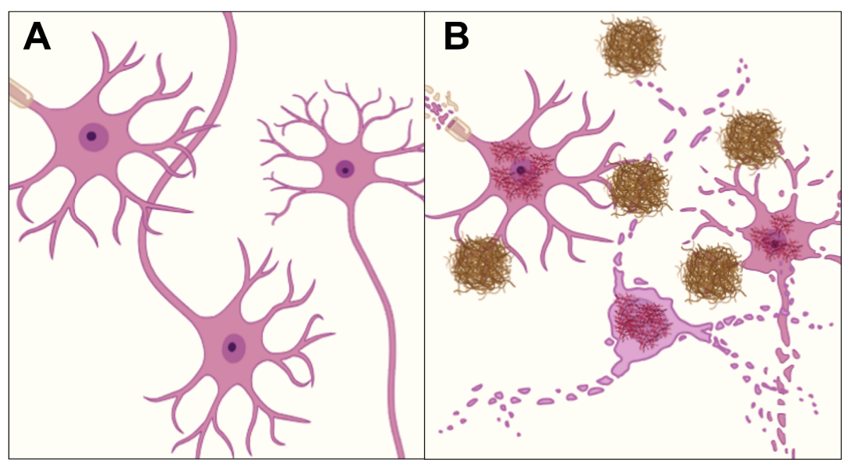
Today, there are thousands of papers, projects, and presentations that use the word “amyloid” and are dedicated to solving the diseases of the world, but I wonder if they know of Nicholas Fontanus or Rudolf Virchow and the mistake that lead us to the world we research today. It is a reminder that one little discovery, even if it isn’t perfect, can change the future of research forever.
References
Annabelle Slingerland. Nicolaas Fontanus, eminent Dutch physician. (2026).
Krystal, H. L., Ross, D. A. & Mecca, A. P. Amyloid: From Starch to Finish. Biol. Psychiatry 87, e23–e24 (2020).
Kyle, R. A. Amyloidosis: a convoluted story. Br. J. Haematol. 114, 529–538 (2001).
Turtz, M., Bone, B. M., Ramos, A. & Dobbs, M. Amyloidosis and Amyloid-Related Diseases: Political Contexts of Early Discoveries. Academic Medicine & Surgery https://doi.org/10.62186/001c.144699 (2025) doi:10.62186/001c.144699.
Schultz, M. Rudolf Virchow. Emerg. Infect. Dis. 14, 1480–1481 (2008).
Westermark, G. T., Johnson, K. H. & Westermark, P. Staining methods for identification of amyloid in tissue. in Methods in Enzymology vol. 309 3–25 (Academic Press, 1999).
Hiramatsu, T. et al. Iodine staining as a useful probe for distinguishing insulin amyloid polymorphs. Sci. Rep. 10, 16741 (2020).
Aterman, K. A historical note on the iodine-sulphuric acid reaction of amyloid. Histochem 49, 131–143 (1976).
DuPre, N. C. et al. Corpora amylacea in prostatectomy tissue and associations with molecular, histological, and lifestyle factors. Prostate 78, 1172–1180 (2018).
Rambaran, R. N. & Serpell, L. C. Amyloid fibrils. Prion 2, 112–117 (2008).
Friedrich, N., Kékulé, A. & Virchow, R. Zur amyloidfrage. in Archiv für Pathologische Anatomie und Physiologie und für Klinische Medizin vol. 16 50–65 (Georg Reimer Verlag, Berlin, 1859).
Sipe, J. D. & Cohen, A. S. Review: History of the amyloid fibril. J. Struct. Biol. 130, 88–98 (2000).
Cohen, A. S. General introduction and a brief history of amyloidosis. in Amyloidosis 3–19 (Springer Netherlands, Dordrecht, 1986). doi:10.1007/978-94-009-4309-4_1.
Nathan Scott Smith. Protein aggregation and drug discovery for alzheimer’s disease and cancer. (Rensselaer Polytechnic Institute, Troy, 2025).